
MicroCT Imaging of Metamorphosis in the African Clawed Toad
MicroCT imaging is an exceptional tool for 3D visualisation. A new Data Note published in GigaScience reports on a unique, high-quality series of MicroCT images that covers the lifespan of an amphibian from tadpole to froglet to mature adult frog. The Data Note reports on a 3D anatomical atlas of an important model organism of […]
The post MicroCT Imaging of Metamorphosis in the African Clawed Toad appeared first on GigaBlog.
MicroCT imaging is an exceptional tool for 3D visualisation. A new Data Note published in GigaScience reports on a unique, high-quality series of MicroCT images that covers the lifespan of an amphibian from tadpole to froglet to mature adult frog. The Data Note reports on a 3D anatomical atlas of an important model organism of development, the African Clawed Toad Xenopus laevis.
The African Clawed Toad as a Model of Development
Xenopus laevis has played an important role in our understanding of embryonic development. In the early twentieth century, Hans Spemann and Hilde Mangold used transplantation experiments in Xenopus laevis embryos to showcase that the fate of cells was dependent on their location.
Xenopus laevis was also used in the early studies of nuclear transplantation and cloning. In 1958, John Gurdon cloned a sexually mature Xenopus laevis toad by transferring a nucleus from the somatic cells of a Xenopus laevis tadpole into an unfertilised Xenopus laevis egg.
Despite the immense understanding of Xenopus early development, there is a gap in our knowledge for the more advanced stages of development from premetamorphosis tadpole to adult. As Dr. Jakub Harnos from Masaryk University (Czech Republic), a lead scientist of the GigaScience study, explained:
“Xenopus laevis, the African clawed frog, is a versatile vertebrate model organism in various biological disciplines, prominently in developmental biology to study body plan reorganization during metamorphosis. However, a notable gap exists in the availability of comprehensive datasets encompassing Xenopus’ late developmental stages.”
MicroCT Imaging of Xenopus laevis
Towards this end, the authors used micro-computed tomography (MicroCT), a non-invasive 3D imaging technique, to more accurately describe Xenopus laevis development. A key advantage of MicroCT is micrometre-scale resolution. In GigaScience we have previously published similar 3D-imaging datasets of the proteus, a mysterious cave-dwelling salamander, to reveal adaptations for life in the dark (see blog).
X-rays used in MicroCT imaging are ideally suited for visualising skeletal components. In addition, through the use of contrast agents, MicroCT can also be used to visualise soft tissues, such as the brain and alimentary system.
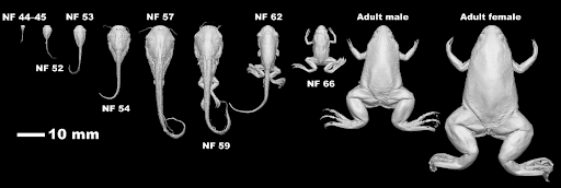
The Xenopus laevis stages of development were carefully chosen with reference to specific morphological structures, such as the presence and dimensions of legs and the tail, or rearrangements of the head.
By capturing development at multiple stages, the authors have created an anatomical atlas, which is an immensely useful spatiotemporal reference framework that enables researchers to more accurately understand developmental dynamics.
MicroCT was used to generate an anatomical atlas of the African clawed frog (Xenopus laevis).
To illustrate the utility of this 3D imaging resource, the authors highlight salient changes that were observed during metamorphosis, and during sexual differentiation.
Visualising Metamorphosis using MicroCT Imaging
Through detailed analysis of the 3D reconstructions at the various stages of development, the authors highlighted that it was possible to pinpoint key changes that are occur in Xenopus laevis from premetamorphosis, prometamorphosis, and climax metamorphosis stages, through to froglet and mature adult stages.
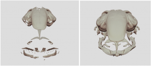
3D reconstructions of developing skull of Xenopus laevis from MicroCT imaging.
There were notable changes in the morphology of the head, as determined by morphometric analysis at multiple stages of development. Most striking was a progressive decrease in eye distance with advancing development. As the authors explained:
“These changes actually occurred in Xenopus in two phases: a gradual increase in the pre-metamorphic stages followed by a peak at the onset of climax metamorphosis and finally, a decrease to a compact head configuration in froglets and adult frogs”.
The authors additionally speculate on the evolutionary implications of developmental trajectory:
“This adaptation aligns well with the frog’s life strategy, transitioning from a water-dwelling tadpole with lateral eyes to an adult with eyes positioned on top of the head for a submerged lifestyle, reminiscent of crocodilians”.
There were additional changes in the morphology of the alimentary system. As the authors state:
These observations on gut coiling morphogenesis, as captured by MicroCT imaging, would be very difficult to study by other means.
Sexual Dimorphism in Xenopus laevis
Sexual dimorphism, which is the phenomenon where the different sexes of a species exhibit different morphological characteristics, is particularly prominent in adult Xenopus laevis, where the female is significantly larger than the male.
Through morphometric analysis of the 3D volumetric models, the authors were able to compare growth patterns. As the authors explained:
“Based on our micro-CT analysis, in terms of sex, we found that adult females can be viewed as essentially enlarged males, indicating a proportional growth pattern”.
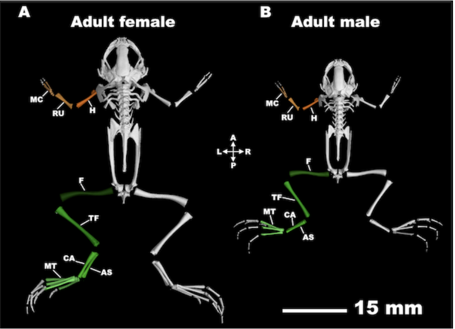
Sexual dimorphism is particularly prominent in adult Xenopus laevis, where the female is significantly larger than the male.
The authors report that size difference is related to sex-related differences in growth of the cartilaginous skeleton. The skeleton first develops as cartilaginous condensations prior to calcification into bone. As the authors explained:
“After climax metamorphosis, no proliferative ossification occurs, and the process is limited to the calcification of cartilage, irrespective of the frog’s sex.”
Hidden Anatomy: Toad’s Teeth
The authors additionally highlighted how 3D imaging was used to uncover morphological detail in anatomical components that were otherwise quite difficult to visualise. The authors used craniofacial development, and specifically teeth patterning, as an illustrative example. As the authors say on this:
“Upon initial observation, the presence of teeth in X. laevis may not be readily apparent.”
The authors further explaining:
“This is due to the fact that teeth were exclusively found in the maxilla, referred to as maxillary teeth and behind the maxillary arch on the vomeral bone – so-called vomeral teeth… In contrast, the mandible contains no teeth”.
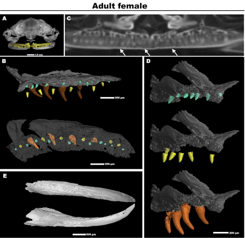
Morphometric analysis of teeth in 3D reconstructions of adult Xenopus laevis.
At GigaScience, we wonder if citizen scientists will uncover additional hidden anatomical components in the Xenopus laevis 3D anatomical atlas.
3D Printable Models of Xenopus laevis
At GigaScience, we see immense value in this 3D anatomical atlas as a spatiotemporal framework to understand vertebrate development . To enable researchers, and also the 3D modelling and 3D printing community to easily access printable 3D models, we have generated a collection of 40 surface-rendered 3D models from the Xenopus laevis anatomical atlas at multiple stages of development. These models can be interactively explore using SketchFab, and the set of 3D printable models are additionally available from the 3D printing community repository Thingiverse.
There is a collection of 30 SketchFab models of Xenopus laevis development, where the sex is not specified, that includes interactive models of various anatomical components.
In addition, there is a collection of 5 SketchFab models of Xenopus laevis adult male, and a collection of 5 SketchFab models of Xenopus laevis adult female, that includes interactive models of the skeletal system, brain, and alimentary system. With examples embedded in the paper using our Sketchfab integration.
For a step-by-step guide on how to 3D print the data from a scientific publication such as this see our previous Blog and video.
The MicroCT data (8-bit TIFF format) 3D reconstructions (STL format) are available for download from Zenodo. Supporting metadata and movie files are additionally available from GigaDB.
References
Laznovsky J; Kavkova M; Reis A; Robovska-Havelkova P; Maia LA; Krivanek J; Zikmund T; Kaiser J; Buchtova M; Harnos J (2024). Unveiling Vertebrate Development Dynamics in Frog Xenopus laevis using Micro-CT Imaging. GigaScience giae037 https://doi.org/10.1093/gigascience/giae037
The post MicroCT Imaging of Metamorphosis in the African Clawed Toad appeared first on GigaBlog.